Sensory system
Introduction
All asteroids lack a centralised nervous system. Instead these organisms possess a nerve ring, surrounding the stomach. Radiating out from this central ring are radial nerves running down the ambulacra of each leg (Ruppert et al. 2004).
This nervous system is broken down into two intra-epithelial nets, the ectoneural system, which is the sensory component of the system, and the hyponeural system, comprising the motor system (Ruppert et al. 2004). These systems are presentin different locations within the starfish, the ectoneural in the epidermis and the hyponeural in the coelomic lining, and are connected by neurons crossing the dermis (Ruppert et al. 2004).
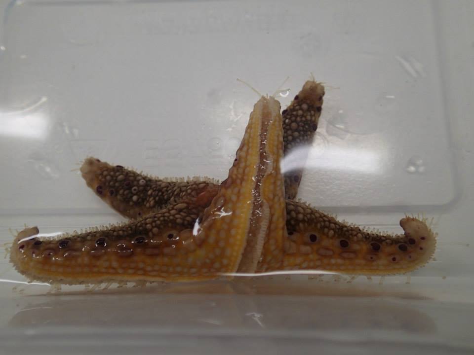
The organs essential for sensory capabilities are the optic cushion (eye spot), tube feet and nerves (Ullrich-Luter et al. 2011). However, when examining N.cumingi, it was observed that the tip of the individuals’ legs was constantly curled upward, suggesting this area possesses sensory structures. Upon closer examination it became apparent that each of these legs possessed tube feet like structures, which lacked a sucker and possessed a darker pigment. These structures were deemed modified tube feet. Although the sensory capabilities of tube feet is known (Ruppert et al. 2004) the role of these modified tube feet has not been documented. As this area is continually turned upward it is hypothesised that these structures possess sensory capabilities. Therefore, this study is aimed at determining the chemical sensory capabilities of these modified tube feet in addition to examining the anatomy of the structure.

The modified tube feet and eye spot of N.cumingi
Methods
In order to test the sensory capabilities of the modified tube feet, numerous stimuli were utilised. These included extracts from the following organisms; sponges, crustaceans, coral, algae, fish and polychaetes. Each of these organisms, with the exception of the sponge and algae, were dead upon collection from the rubble, broken up a few days prior, or from the plankton tow, in the case of the fish. These organisms were then crushed using a mortar and pestle until liquid was produced. Testing commenced when the sea star situated itself into the experimental position (figure 1), which the individual moved into naturally. Once in this position, the modified tube feet present on the leg facing upward were stimulated by placing two drops of the liquid extracted from each organism into the water approximately one centimetre away from the modified tube feet, one extract at a time. The response was filmed and recorded as either no response or response. Once completed, the starfish was removed and the water changed. Experimentation commenced once again when the starfish was in the experimental position.
Upon completion of experimentation, four of the five tips of the legs were removed with the use of a scalpel. Prior to this, the starfish was placed in the freezer for a 20-minute period in order to minimize pain. These feet were then placed into a fixation agent (4% PFA) for two hours at which point they were removed and placed into 10mM of EDTA. These leg tips were contained within EDTA for a two week period, until the structures were soft enough to be sectioned. After two weeks in EDTA, the leg tips were placed into a solution of 70% ethanol ready for sectioning. The legs were sectioned from the tip to the first red spot, present on the sides of each leg.

figure 1. the experimental position of N.cumingi
Results
The tube feet appeared to respond to only one stimulus, the Amphimedon queenslandica (sponge) extract. Upon connection with the extract the feet immediately retracted, and the entire leg began to curl back, away from the sponge extract. However, when in contact with the coral, algae, polychaete and fish the feet did not respond at all. These responses can be seen in the video below.
Table 1. Response of modified tube feet of N.cumingi to a variety of stimuli
|
Organism
|
Response of modified tube feet
|
|
Coral
|
No response
|
|
algae
|
No response
|
|
polychaete
|
No response
|
|
fish
|
No response
|
|
sponge
|
Response
|
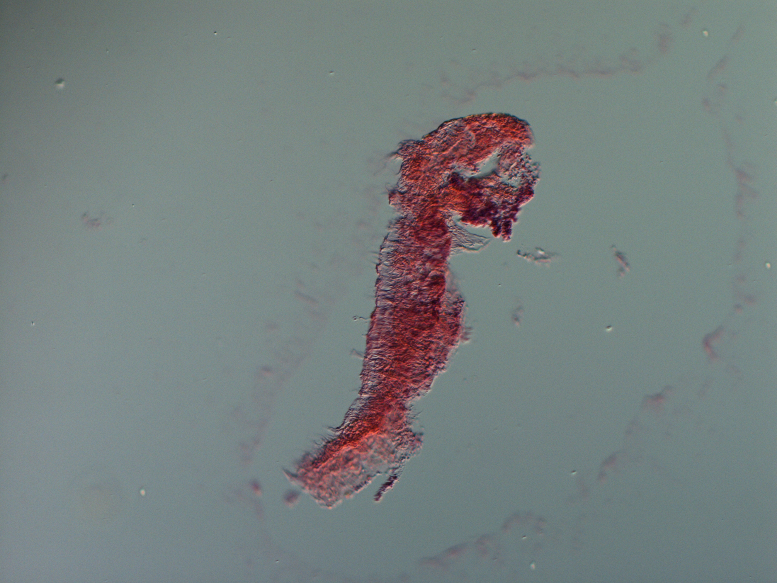
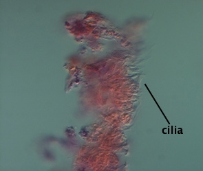
The sections produced from the tips of the starfish feet displayed a number of structures. One of these structures was clearly not the leg itself, as it did not possess the correct shape, and was hypothesised to be a cross section of a sensory tube foot. This structure was long and cylindrical in shape and possessed high densities of muscle fibres (figure 2). The primary feature identified on this structure was cilia (figure 3).
 
figure 2. The section of a modified tube foot of N.cumingi
 
figure 3. section of N.cumingi modified tube foot demonstrating the cilia present on the structure
Discussion
The primary aim of this study was to examine the role of the modified tube feet and to determine whether they, in fact, play a role in the sensory system, as hypothesised.
The results of the stimulation experiment suggest that these modified tube feet possess sensory capabilities, as the structure demonstrated a clear negative response to the sponge extract. As sponges are often toxic, and those living closer to the equator generally possess higher toxicity (Green 1977), it is possible that Amphimedon queenslandica is toxic. These modifiedtube feet may therefore be sensing this toxicity, which may explain their retraction.
Additionally, cilia are important aspects of the sensory system, present in the rods and cones within the eyes, the hairs of ears and the olfactory neuron receptors (Perkins et al. 1986). This suggests that the presence of cilia on these tube feet is further support for the hypothesis that these structures possess sensory capabilities. It is therefore speculated that these cilia present on the modified tube feet may be responsible for the retraction of the leg and feet in response to A.queenslandica.
As these structures could be accountable for environmental chemical sensing, they may be useful in detecting potential prey items, as this is accomplished via chemotaxis (Whittle & Blumer 1970) or may play a role in the orientation of starfish movement, which was also determined to be via chemosensory capabilities (Dale 1999).
Future studies expanding on the stimulation experiment need to be undertaken with a larger sample size and further replication, in order to test the robustness of the results. |